SHRO’s Giordano to Speak at Virtual Workshop on Artificial Intelligence in Medicine
- Post & News
- admin
- March 7, 2023
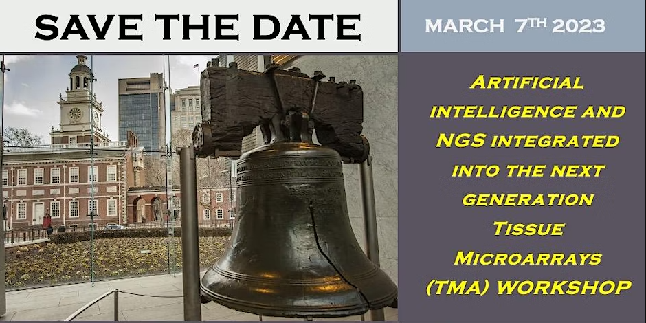
Artificial Intelligence and NGS Integrated into the Next Generation Tissue Microarrays (TMA) Workshop
Originally published in Newswise

Newswise — March 7, 2023, Philadelphia – Antonio Giordano, M.D., Ph.D., Founder and President of the Sbarro Health Research Organization (SHRO), is scheduled to speak at a virtual workshop on Artificial Intelligence (AI) in medicine.
Event Details
7TH IN PERSON or VIRTUAL WORKSHOP ON “Artificial intelligence and NGS integrated into the next generation Tissue Microarrays (TMA)”
MARCH 7TH 2023, 9 AM -5 PM
DON’T MISS THIS OPPORTUNITY TO LEARN MORE ABOUT Artificial intelligence and NGS integrated into the next generation Tissue Microarrays
PLEAS REGISTER AT THE LINK: https://lnkd.in/ennhsFsW
The workshop will focus on AI and next-generation tissue microarrays (TMA). TMA is a versatile technique with definite clinical and pathological applications. Originally was used in cancer research however its applications are rapidly expanding to non-neoplastic diseases: neurodegenerative, dermatological, cardiac and placental disorders. The new generation of tissue microarrays looks at the benefits of coupling digital pathology in translational research to deliver more efficient and time effective solutions to answer complex clinical and scientific questions. At the core of next generation TMAs is digital pathology and artificial intelligence which allows the identification of specific areas where to focus and better target investigations.
The tissue microarray format provides opportunities for digital imaging acquisition, image processing and database integration. Advances in digital imaging help to alleviate previous bottlenecks in the research pipeline, permit computer image scoring and convey telepathology opportunities for remote image analysis. In addition, the TMA technology gives the possibility to extract near by Tissue Cores which are inserted in the TMA block and in a mictotile plate. This gives the opportunity to ectract DNA/RNA form the tissue cores in the microtile plate and perform NGS analysis from the tissue sample. The advantage of these combined technologies is to have the digital image of the TMA core and the genetic anlysys from the same patient/donor.
TMA applications are wide: predictive, to authenticate biomarkers with the objective to annotate treatment/therapy responses; prognostic in order to reduce cancer incidence and mortality; in clinical applications to determine the functional status of the cancer before therapy initiation; (v) in diagnostic pathology to qualify samples, reduce laboratory variations and results interpretation. Moreover, the TMAs formats provide opportunities for digital imaging acquisition, image processing and database integration
